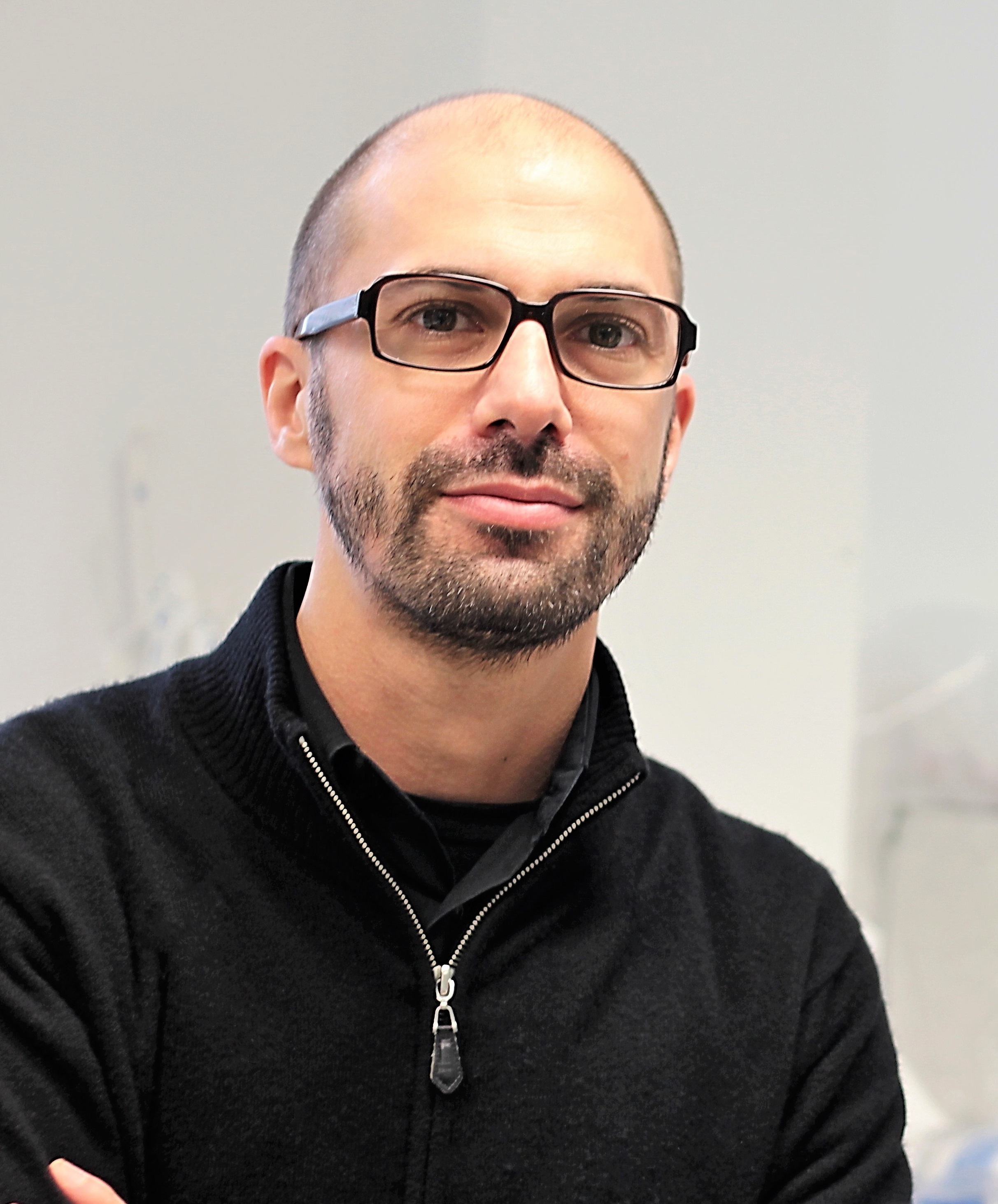
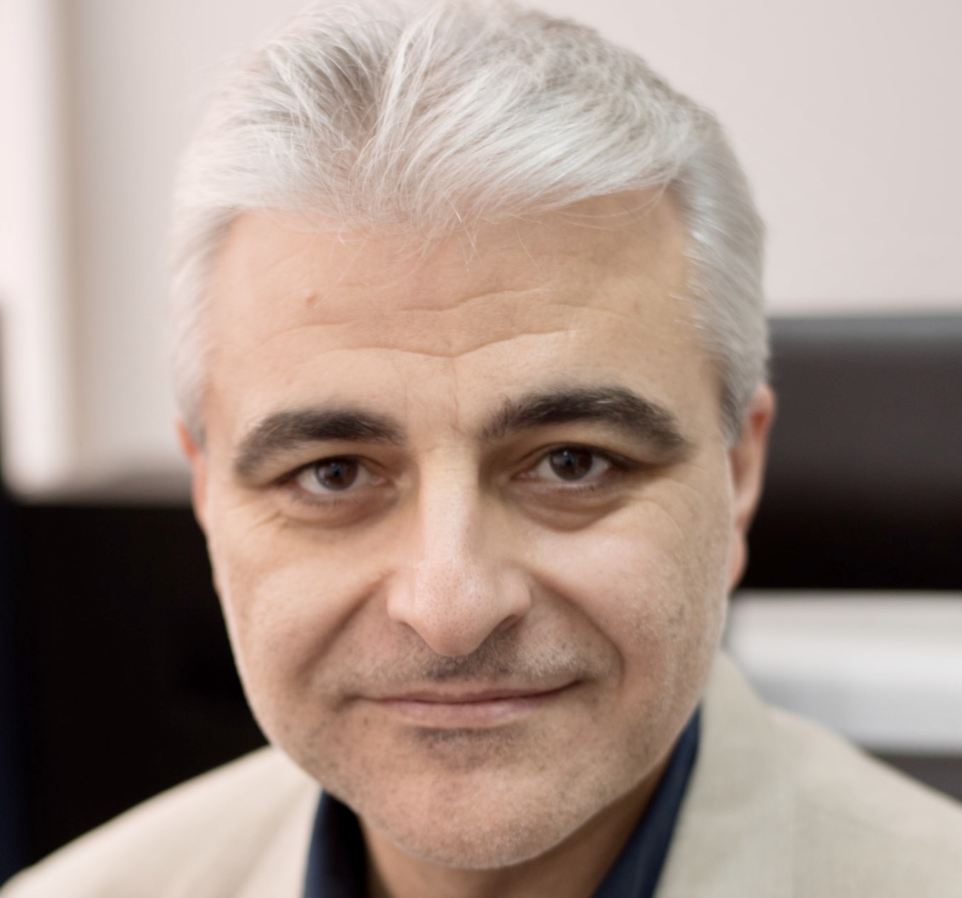
Meet our invited speakers:
- KEYNOTE: Eeva-Liisa Eskelinen (University of Turku, Finland)
Title: Membrane dynamics during autophagosome biogenesis: what correlative light-electron microscopy and 3D electron microscopy can tell us
Abstract: The origin of phagophore and autophagosome membranes is one of the long-lasting questions in autophagy research. Autophagosomes are known to form in close connection with the endoplasmic reticulum (ER) subdomains called omegasomes. In order to elucidate the membrane dynamics during autophagosome biogenesis, we have used live-cell imaging, correlative light-electron microscopy and three-dimensional (3D) electron microscopy to investigate the early steps of phagophore biogenesis in non-selective and selective autophagy in cultured mammalian cell lines. Our results show that both non-selective and selective autophagosomes form in association with the ER. In addition, using cells expressing early phagophore markers, we have captured 3D images of phagophore precursors. The images show that phagophores emerge as small round structures from the ER, with additional membrane contact sites with other membranous organelles. We observe clusters of small, often cup-shaped vesicles adjacent to the phagophore precursors. At a later stage the phagophores have multiple contact sites with the ER, and we also see membrane contact sites with mitochondria and vesicles of different sizes.
Model systems: cultured mammalian cell lines

- Christian Behrends (Ludwig-Maximilians-Universität (LMU), Germany)
Title: Exploring machinery and cargo of selective autophagy
Abstract: Lysosomes represent the terminal subcellular compartment for a number of degradative and recycling pathways which makes their integrity crucial for cell homeostasis. However, under certain stress and pathophysiological conditions (e.g. neurodegeneration) the lysosomal membrane can be damaged resulting in functional impairment, leakage of lysosomal content (e.g. protons, reactive oxygen species and degradative enzymes) into the cytosol and eventually lysosomal cell death. To cope with lysosomal damage the cell therefore developed several defense strategies which are summarized under the term endo-lysosomal damage response. Selective autophagy of the ruptured lysosome, termed lysophagy, presents the ultimate step for incurable damage and is accompanied by a fast and robust ubiquitylation of the lysosome. We are particularly interested in identifying the ubiquitylation machinery and their substrates upon lysosomal damage.
Model systems: cultured mammalian cell lines

- Ian Ganley (University of Dundee, UK)
Title: Strengthening the link between Parkinson’s disease and mitophagy
Abstract: Parkinson’s disease (PD) is a progressive neurodegenerative disorder that is characterised by the loss of midbrain dopaminergic neurons. The physiological and molecular mechanisms leading to this neuronal loss are currently unclear; however, mitochondrial and lysosomal dysfunction seem to play a central disease role. This is highlighted by the fact that PINK1 and Parkin, two proteins that can be mutated in familial PD, play a key role in initiating mitophagy. Using mouse models to study mitophagy and autophagy in vivo, we have found that mitophagy is a prevalent physiological process, yet PINK1 and Parkin-dependent mitophagy only accounts for a fraction of this. What about these other pathways – could they also be relevant in PD? Here I will describe our recent work exploring if the most common mutation found in PD, LRRK2 G2019S, also impacts mitophagy.
Model systems: in vivo mouse models, cultured mammalian cell lines

- Etienne Morel (Institut Necker Enfants Malades, France)
Title: Autophagy machinery mobilization in ciliogenesis during nutritional stress: a non-canonical response
Abstract: The primary cilium (PC), a plasma membrane microtubule-based structure, is a sensor of extracellular chemical and mechanical stress stimuli. Upon ciliogenesis, the autophagy protein ATG16L1 and the ciliary protein IFT20 are co-transported to the PC. In a recent study we demonstrated, in kidney polarized epithelial cells, that IFT20 and ATG16L1 interact in a multiprotein complex. This interaction is mediated by the ATG16L1 WD40 domain and an ATG16L1-binding motif newly identified in IFT20. ATG16L1-deficient cells are decorated by giant ciliary structures hallmarked by defects in PC-associated signaling. These structures uncommonly accumulate phosphatidylinositol-4,5-bisphosphate (PtdIns[4,5]P2) while phosphatidylinositol-4-phosphate (PtdIns4P), a lipid normally concentrated in the PC, is excluded. We show that INPP5E, a phosphoinositide-associated phosphatase responsible for PtdIns4P generation, is a partner of ATG16L1 in this context. Perturbation of the ATG16L1-IFT20 complex alters INPP5E trafficking and proper function at the ciliary membrane. Altogether, these results reveal a novel autophagy-independent function of ATG16L1 that contributes to proper PC dynamics and function.
Model systems: cultured mammalian cell lines

- Tassula Proikas-Cezanne (University of Tuebingen, Germany)
Title: Regulation of WIPI1 function in autophagy
Abstract: Autophagy is an evolutionary conserved eukaryotic bulk degradation pathway that results in regulated cellular clearance and secures cellular survival. Autophagic dysfunction has been found to be associated with various human diseases including cancer and neurodegeneration. Aiming to develop therapeutics capable to cure age-related human diseases, the molecular details of autophagy are now becoming understood. We identified the human WIPI gene family (WIPI-1, -2, -3, and -4) and have begun to establish that WIPI genes are aberrantly expressed in a variety of human cancers (Proikas-Cezanne et al., Oncogene 2004). Using biochemical techniques coupled with confocal and electron microscopy we have demonstrated that WIPI-1 (Atg18 in S. cerevisiae) specifically binds PI(3)P at the onset of autophagy and is essential for autophagosome formation. Upon binding to PI(3)P, WIPI-1 protein accumulation at autophagosomal membranes (WIPI-1 puncta-formation) can be visualized by fluorescent microscopy, representing a novel monitoring system for mammalian autophagy. We have started to employ the WIPI-1 puncta-formation assay for automated high content screening purposes and found that this assay is suitable for drug and siRNA screening approaches, as well as for high through-put analyses of mammalian autophagy. Currently, we are assigning a biological function to all of the different human WIPI family members and wish to unravel the signaling network regulating WIPI-meditaed autophagy as well as the WIPI interactome. We aim to target the autophagic activity in human diseases by modulating WIPI gene expression and WIPI protein activity.
Model systems: Primary mouse and human cells, human tumor cell lines, C. elegans, M. musculus (in cooperations), S. cerevisiae (in cooperations), human iPS (in cooperation).

- Patricia Boya (Spanish National Research Council (CSIC), Spain)
Title: Selective autophagy plays a protective role against acute and age-related retinal degeneration
Abstract: Age-related macular degeneration (AMD) is the leading cause of blindness in elderly people in the developed world, and the number of people affected is expected to almost double by 2040. The retina presents one of the highest metabolic demands in our body that is partially or fully fulfilled by mitochondria metabolism. Together with its post-mitotic status, this context requires a tightly-regulated housekeeping system that includes selective mitochondrial autophagy. We want to assess the effects of selective autophagy deficit or induction in the retina given an AMD-like paradigm.
Ambra1+/gt autophagy-deficient mice present alterations in the RPE, similar to those observed in human AMD patients, which appear in an age-dependent manner. Furthermore, Ambra1+/gt mice are more sensitive to acute sodium iodate induced retinal degeneration than their Ambra+/+ littermates. Our data also show that Urolithin A induced mitophagy in vivo and prevented neurodegeneration both in the neuroretina and RPE. This amelioration was also associated with decreased lipid peroxidation, gliosis and increased photoreceptor survival. In vitro, inhibition of mitophagy, or general macroautophagy, abolished this rescue. In conclusion our data show that selective autophagy plays a protective role in the retina and can be exploited to preserve vision in physiological or pathological conditions.
Model systems: cultured mammalian cell lines, in vivo mouse models

- Nektarios Tavernarakis (Foundation for Research and Technology-Hellas (FORTH), Greece)
Title: Nuclear autophagy delays ageing and preserves germline immortality under stress
Abstract: The crosstalk between autophagy and the nucleus in the context of physiology and pathology remains largely elusive. We investigated this potential association by dissecting the involvement of nuclear membrane components in the autophagic process. To this end, we examined the role of the highly conserved, C. elegans nuclear envelope anchorage protein 1 (ANC-1) and its mammalian orthologues Nesprin 1 and 2 in autophagy. These are large, multi-domain, outer nuclear membrane proteins that maintain nuclear integrity. Nesprin 1 and 2 isoforms are highly abundant in multiple tissues, including the brain, heart, liver and kidney. In humans, polymorphisms in Nesprin 1 and 2 have been implicated in neurodevelopmental disorders, such as learning disability, autism and ataxia, as well as, in muscular dystrophies, in cancer and ageing. Using both C. elegans and mouse models we uncovered a novel link between these nuclear envelope components and autophagic processes. We find that autophagy regulates Nesprin 1 and 2 protein levels under both basal conditions and nutrient stress. Conversely, both C. elegans ANC-1 and mouse Nesprins interact with, and regulate the abundance and localization of general and selective autophagic machinery components such as LGG-1/LC3B and SQST-1/p62. Notably, these nuclear envelope components act downstream of autophagy under nutrient stress to maintain nucleolar shape and integrity. Thus, cross-regulation between autophagy and ANC-1/Nesprin proteins promotes nuclear homeostasis and safeguards the nucleolus and nuclear proteins under stress and during ageing.
Model systems: C. elegans
