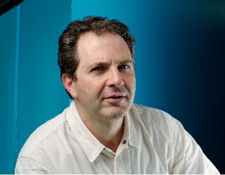
KEYNOTE 1: Katja Simon (katja.simon@mdc-berlin.de; Max-Delbrück-Centrum für Molekulare Medizin ; Berlin, Germany)

Title: Autophagy’s role in immune aging
Abstract: Across all arms of the immune system, the process of autophagy is key in development, function, prevention of senescence and homeostasis. Particularly long lived cells such as stem cells require autophagy for maintenance as they tend to divide infrequently. However, the mechanisms involved are much less clear, and encompass control of metabolism, cell survival, and selective degradation of substrates and organelles. Autophagy’s contribution to this process is for example the maintenance of mitochondrial quality. While so far, the autophagy field has focused on degradation rather than provision, we found in hematopoietic stem cells that balancing the provision of amino acids by autophagy versus importing them is key to their maintenance. In T and B lymphocytes autophagy keeps the cells young, and markers of senescence can be reversed by replenishing essential metabolites such as spermidine that are needed for autophagy. Our work is based on mouse models, human blood cells and clinical trials. The most commonly used techniques include multispectral flow cytometry, proteomics, metabolomics and scRNA. I will summarise our data on autophagy’s impact on the immune system, with a particular emphasis on differentiation, maintenance and aging in mouse and human.
Model systems: mouse models, human blood cells and clinical trials
KEYNOTE 2: Anne Simonsen (a.g.simonsen@medisin.uio.no; University of Oslo, Norway)

Title: Exploring the crosstalk of hypoxia-induced mitophagy and cellular bioenergetics
Abstract: A healthy mitochondrial population is maintained through a series of quality control pathways and a fine-tuned balance between mitochondrial biogenesis and degradation. Disruption of this delicate balance leads to mitochondrial dysfunction and contributes to aging and several diseases such as neurodegeneration, cardiac disease and cancer. Mitophagy involves lysosomal degradation of mitochondrial components and can be induced by a variety of stress and damage stimuli, including hypoxic conditions. However, our mechanistic understanding of mitophagy is only just emerging, and there are currently no therapies targeting mitochondrial quality control. We have recently identified several novel regulators of hypoxia-induced mitophagy using high throughput siRNA screens in mammalian cells expressing mitophagy reporters as well as different omics approaches. I will here present data showing that novel regulators of mitophagy control cellular bioenergetics and disease development in zebrafish models.
Model systems: cultured mammalian cell lines; zebrafish models
Christian Münz (christian.muenz@uzh.ch; University of Zürich, Switzerland)

Title: Autophagy during human tumorvirus infections
Abstract: Autophagy serves as a defense mechanism against intracellular pathogens. The escape of pathogens from endosomes is closely monitored for membrane damage by the autophagic machinery. We could demonstrate that lipidation of the autophagy protein LC3 is induced shortly after entry of the human oncogenic Kaposi sarcoma associated herpesvirus (KSHV). Both nuclear dot protein 52 kDa (NDP52), a receptor of selective autophagy, and the endolysosomal damage sensor galectin-8 were recruited to endocytosed viral particles. Loss of components in the LC3 lipidation complex, NDP52 or galactin-8 increased infection. Using herpes simplex virus, influenza virus, listeriolysin, and adenovirus as negative and positive controls, and in contrast to what was previously thought about enveloped viruses, KSHV binding to EphA2 by its envelope protein gH caused endolysosomal membrane damage, akin to non-enveloped viruses and bacteria. This results in KSHV entry restriction by autophagy. In contrast to this antiviral function of the autophagic machinery certain herpesviruses include autophagic membranes into their infectious virus particles. We found several components of the autophagy machinery, including membrane-associated LC3B-II, in virions of the human oncogenic Epstein Barr virus (EBV). Additionally, we showed that viral capsid assembly proteins BVRF2 and BdRF1 interact with LC3B-II via their common protein domain. Using an EBV mutant, we identified BVRF2 as essential to assemble mature capsids and produce infectious EBV. However, BdRF1 was sufficient for the release of non-infectious viral envelopes as long as autophagy was not compromised. These data suggests that BVRF2 and BdRF1 are critical for EBV envelope release by interacting with the autophagic machinery.
Model systems: cultured mammalian cell lines
Joern Dengjel (joern.dengjel@unifr.ch; University of Fribourg, Switzerland)

Title: Phosphorylation-based regulation of autophagy
Abstract: Autophagy initiation is regulated on a posttranslational level involving posttranslational modifications of proteins that affect protein activity and subcellular localization. Protein and lipid kinases and phosphatases play pivotal roles in autophagy induction. To gain new insights into autophagy induction, we generated a deep ULK1 complex interactome by combining affinity purification- and proximity labelling-mass spectrometry of all four ULK1 complex members: ULK1, ATG13, ATG101 and RB1CC1/FIP200. Under starvation conditions, the ULK1 complex interacts with several protein and lipid kinases and phosphatases implying the formation of a signalosome. Interestingly, also several selective autophagy receptors interact with ULK1 indicating the activation of selective autophagy pathways by nutrient starvation. In the current project, we characterize new protein-protein interactions and posttranslational modifications important for functional autophagy addressing amongst others the subcellular localization of protein complexes. We highlight the interplay of ULK1 with the HSP70 co-chaperone BAG2 and how BAG2 influences AMBRA1 localization and with this activity of the VPS34 lipid kinase complex. In addition, we characterize new lipid binding proteins which influence autophagosome biogenesis. Our data highlight the robustness of autophagy initiation and the complexity of underlying signaling pathways.
Model systems: cultured mammalian cell lines
Jayanta Debnath (Jayanta.Debnath@ucsf.edu; University of California San Francisco, USA)

Title: Autophagy suppresses pro-metastatic differentiation by degrading SQSTM1-NBR1 condensates
Abstract: Although autophagy inhibitors are widely being repurposed for cancer treatment in clinical trials, our past work unexpectedly uncovered that autophagy suppresses metastatic recurrence and outgrowth in both mouse and human breast cancer models. Our results now demonstrate that autophagy normally suppresses breast cancer metastasis by enabling the clearance of NBR1-p62/SQSTM1 condensates that instruct p63-mediated pro-metastatic basal differentiation programs. When autophagy is inhibited, the autophagy cargo receptors NBR1 and p62/SQSTM1 accumulate within biomolecular condensates in cells, which drives basal differentiation in mouse and human breast cancer models. Accordingly, a mutant form of NBR1 (NBR1-D50R) unable to bind p62/SQSTM1 is sufficient to disrupt condensate formation, which prevents basal differentiation and abrogates metastasis in vivo. Mechanistically, these NBR1-p62/SQSTM1 condensates activate p63 by sequestering ITCH, an ubiquitin ligase that degrades and negatively regulates p63 in breast cancer cells. Furthermore, pharmacological autophagy inhibition in primary organoids generated from breast cancer patients and those at high-risk for breast cancer triggers both condensate formation and pro-metastatic basal differentiation programs. Overall, our findings illuminate how proteostatic defects arising in the setting of therapeutic autophagy inhibition promote metastatic progression.
Model systems: mouse and human breast cancer models
Maho Hamasaki (hamasaki@fbs.osaka-u.ac.jp; Osaka University, Japan)

Title: Palmitoylation of ULK1 by ZDHHC13 plays a crucial role in autophagy
Abstract: Autophagy is a highly conserved process from yeast to mammals in which intracellular materials are engulfed by a double-membrane organelle called autophagosome and degrading materials by fusing with the lysosome. The process of autophagy is regulated by sequential recruitment and function of autophagy-related (Atg) proteins. Genetic hierarchical analyses show that the ULK1 complex comprised of ULK1-FIP200-ATG13-ATG101 translocating from the cytosol to autophagosome formation sites as a most upstream ATG factor; this translocation is critical in autophagy initiation. However, how this translocation occurs remains unclear. Here, we show that ULK1 is palmitoylated by palmitoyltransferase ZDHHC13 and translocated to the autophagosome formation site upon autophagy induction. We find that the ULK1 palmitoylation is required for autophagy initiation. Moreover, the ULK1 palmitoylated enhances the phosphorylation of ATG14L, which is required for activating PI3-Kinase and producing phosphatidylinositol 3-phosphate, one of the autophagosome membrane's lipids. Our results reveal how the most upstream ULK1 complex translocates to the autophagosome formation sites during autophagy.
Model systems: cultured mammalian cell lines
Oliver Florey (Oliver.Florey@babraham.ac.uk; Babraham Institute, Cambridge, UK)

Title: New roles for non-canonical autophagy in lysosome homeostasis
Abstract: Lysosomal damage is a common and severe stress condition that is relevant for degenerative disease, infection, cancer and ageing. Maintaining the integrity of the lysosomal system is therefore of paramount importance for cellular homeostasis. Our previous work has uncovered a non-canonical autophagy pathway that directs the Conjugation of ATG8 proteins to Single Membranes (CASM) upon ionic and pH perturbation of endolysosomal compartments. This is distinct from the canonical autophagy pathway associated with autophagosome formation. We have elucidated the upstream molecular mechanisms regulating the initiation of CASM and identified a wide variety of stimuli that activate it. Our new work now reveals that CASM is a rapid response pathway to lysosome damage/stress and intersects with signalling pathways involved in the repair, biogenesis, turnover and dynamic remodeling of the lysosomal system.
Research Summary: Our lab uses microscopy and cell biology to study the molecular mechanisms involved in maintaining lysosome homeostasis during stress associated with ageing and disease.
Model systems: cultured mammalian cell lines
Sharon Tooze (Sharon.Tooze@crick.ac.uk; The Francis Crick Institute, London, UK)

Title: WIPI2b recruitment to phagophores and ATG16L1 binding are regulated by ULK1 phosphorylation
Abstract: One of the key events in autophagy is the formation of a double-membrane phagophore, and many regulatory mechanisms underpinning this remain under investigation. WIPI2b is among the first proteins to be recruited to the phagophore and is essential for stimulating autophagy flux by recruiting the ATG12~ATG5-ATG16L1 complex, driving LC3 and GABARAP lipidation. Here, we set out to investigate how WIPI2b function at this stage in autophagosome biogenesis is regulated. Based on the interaction of WIPI2 with ULK1, and changes in the migration of WIPI2 by SDS-PAGE we hypothesized WIPI2 phosphorylated, and phosphorylation is frequentently implicated in regulation of function. We studied two phosphorylation sites on WIPI2b, S68 and S284. Phosphorylation at these sites play two distinct roles – regulating WIPI2b's association with ATG16L1 and the phagophore, respectively. Phosphorylation at S68 disrupts ATG16L1 binding. We confirm WIPI2b is a novel ULK1 substrate, and validated this by detection of endogenous phosphorylation at S284. Notably, S284 is situated within an 18-amino-acid stretch, which, when in contact with liposomes forms an amphipathic helix. Phosphorylation at S284 disrupts the formation of the amphipathic helix, hindering the association of WIPI2b with membranes and autophagosome formation. Understanding these intricacies in the regulatory mechanisms governing WIPI2b's association with its interacting partners and membranes, holds the potential to shed light on these complex processes, integral to phagophore biogenesis.
Model systems: cultured mammalian cell lines
Terje Johansen (terje.johansen@uit.no; The Artic University of Norway, Norway)

Title: The mitochondrial ubiquitin ligase MUL1 recruits the ULK1 complex to induce mitophagy
Abstract: The clearance of damaged or surplus mitochondria by selective autophagy or mitophagy is important for maintenance of mitochondrial integrity, cellular homeostasis, and prevention of disease. Our current understanding of mitochondrial quality control and mitophagy comes mostly from studies of stress- or damage-induced mitochondrial degradation involving the kinase PINK1 and the E3 ubiquitin protein ligase PARKIN. Here, we show that the mitochondrial SUMO-E3 ubiquitin ligase, MUL1 induces mitophagy in human HeLa cells independent of its E3-ligase activity and independent of PARKIN. MUL1 binds directly to ULK1 to recruit ULK1 to the mitochondria to induce mitophagy.
Model systems: cultured mammalian cell lines
Thierry Galli (thierry.galli@inserm.fr; Institute of Psychiatry and Neuroscience of Paris, INSERM UMR1266/University Paris Cité, Paris, France)

Title: Molecular and Cellular Mechanisms of Late Endosomal Autophagy-Dependent Secretion
Abstract: The vesicular SNARE VAMP7 facilitates the fusion of late endosomes and lysosomes with the plasma membrane, playing a crucial role in the release of exosomes. Recent proteomics analysis of the secretome revealed that VAMP7 mediates the secretion of endoplasmic reticulum (ER) elements and the pro-form of the neuropeptide VGF, a pathway dependent on ATG5, indicating an involvement of autophagy (1–4). Transcriptomics analysis identified alterations in mitochondrial gene expression in VAMP7 knockout (KO) epithelial cells. Further investigations revealed significant morphological and functional mitochondrial defects in VAMP7 KO cells, observed through electron microscopy, confocal microscopy, and respirometry. Our findings indicate that VAMP7 KO cells exhibit impaired transport of mitochondrial-derived vesicles to late endosomes and reduced secretion of mitochondrial components. Similarly, VAMP7 is essential for the transport and secretion of ER elements to late endosomes. Additionally, stress in the ER or mitochondria increases the release of their respective elements in a VAMP7- and ATG5-dependent manner. We conclude that late endosomal secretion is a critical mechanism for the quality control of both the ER and mitochondria, particularly during stress responses, via an autophagy-dependent pathway.
Model systems: cultured mammalian cell lines
Marja Jaatela (mj@cancer.dk; University of Copenhagen, Denmark)

Title: Control of lysosomal ion homeostasis
Abstract: Calcium and pH homeostasis are of crucial importance for virtually all aspects of cellular life. In mammalian cells, lysosomal concentrations of calcium and protons are maintained three to four orders of magnitude higher than in the cytosol, and the proper function of lysosomes depends on their ability to store and release these ions. Here, I will discuss the mechanisms by which lysosomes maintain their ion homeostasis and how this homeostasis can be targeted in cancer cells.
Model systems: cultured mammalian cell lines; mouse models
Francesco Cecconi (cecconi@cancer.dk; (Danish Cancer Society Research Center, Copenhagen, Denmark and Università Cattolica del Sacro Cuore, Rome, Italy)

Title: Novel forms of selective autophagy and cancer ontogenesis and progression
Abstract: In recent years, we identified different forms of selective autophagy targeting centriolar satellites, peroxisomes and micronuclei. Besides providing an overview of our data in the context of the molecular mechanisms of these processes, I will discuss as this novel scenario provides an alternative explanation for metabolic switches occurring in cancer, reorienting research towards developing innovative therapeutic strategies.
Model systems: cultured mammalian cell lines; mouse models