The 12th Microscopy Webinar will take place on October 25th from 16:00 to 17:00 (Swiss time)
Speaker:
Dimitrii Tanese - Institut de la Vision (France)
Title: Light shaping for 2-photon optogenetics and neuronal circuits manipulation
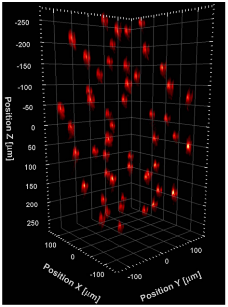
The capability to pattern light deep in biological tissues is crucial for multi-cell optical manipulation and imaging. In particular, in the field of Neuroscience, the advent of novel optogenetic tools, such as light-sensitive channels (opsins) and fluorescent reporters, opened new ways to manipulate and monitor neuronal activity by optical means. The full exploitation of these tools requires the development of optical approaches enabling fast, volumetric, in-depth illumination of multiple targets with single-cell resolution.
Here I will describe a light shaping technique, named Computer Generated Holography, based on the use of liquid crystal spatial light modulator (SLM) to control the phase of a femtosecond infrared laser beam. This approach allows to tailor the illumination profile to target specific subcellular structures or single and multiple cell bodies and to achieve 2-photon optogenetic activation of neurons with single cell resolution deep in scattering tissue[1,2].
I will discuss some potential applications of this approach to investigate neuronal circuits and perform in-vivo synaptic connectivity mapping.
Finally, I will present a recently developed configuration capable of generating sequences of 3D illumination patterns at several kHz rate, overcoming the current limitations of the refresh rate of the SLM liquid crystals. These sequences of multi-spot patterns were exploited either to synchronize spiking activity of pair of cells with sub-millisecond precision or to activate large neuronal ensembles with a reduced power requirement and minimizing potential photoheating[3].
Overall, these approaches opens the way to an all-optical and minimally invasive manipulation of large neuronal circuits, with high spatial and temporal precision, both in-vitro and in-vivo.
References:
[1] Accanto, N., Molinier, C., Tanese, D. et al. (2018); Optica, 5(11), 1478.
[2] Chen, I., et al. (2018); Curr. Opin. Neurobiol. 50, 179-189.
[3] Faini, G., Tanese, D. et al. (2018); BioRrxivV, 448315.